| 1. |
Sun, H., He, Y., Qiao, S. et al. Highly sensitive H2S-LITES sensor with 80 m fiber-coupled multi-pass cell based on optical path multiplexing technology. Photoacoustics, 2025.

|
|
| 2. |
Zha, S., Chen, H., Liu, C. et al. Multivariate-coupled-enhanced photoacoustic spectroscopy with Chebyshev rational fractional-order filtering algorithm for trace CH4 detection. Photoacoustics, 2025.
|
|
| 3. |
Ma, H., Chen, Y., Qiao, S. et al. A high sensitive methane QEPAS sensor based on self-designed trapezoidal-head quartz tuning fork and high power diode laser. Photoacoustics, 2025.
|
|
| 4. |
Hou, J., Liu, X., Sun, H. et al. Dual-Component Gas Sensor Based on Light-Induced Thermoelastic Spectroscopy and Deep Learning. Analytical Chemistry, 2025, 97(9): 5200-5208.
|
|
| 5. |
Hou, J., Liu, X., Sun, H. et al. Dual-Component Gas Sensor Based on Light-Induced Thermoelastic Spectroscopy and Deep Learning. Analytical Chemistry, 2025, 97(9): 5200-5208.
|
|
| 6. |
Ma, H., Qiao, S., He, Y. et al. Load capacitance matching for resonant frequency adjusting-based multi-quartz tuning fork-enhanced laser spectroscopic sensing. Optics Express, 2025, 33(5): 9423-9433.
|
|
| 7. |
Qi, L., Chen, W., Qiao, S. et al. Mid-infrared quasi-distributed carbon monoxide gas sensing based on QEPAS and hollow waveguide. Infrared Physics and Technology, 2025.
|
|
| 8. |
Ma, H., Qiao, S., He, Y. et al. A Highly Sensitive Light-Induced Thermoelastic Spectroscopy Sensor Using a Charge Amplifier to Improve the Signal-to-Noise Ratio. Sensors, 2025, 25(3): 946.
|
|
| 9. |
Fang, F., Wang, R., Shao, D. et al. Improved T-shaped quartz tuning fork with isosceles-trapezoidal grooves optimized for quartz-enhanced photoacoustic spectroscopy. Photoacoustics, 2025.
|
|
| 10. |
Rong, S., Sun, X., Yang, Y. et al. A Trace C2H2 Detection Based on Near-Infrared Dual-Comb Spectroscopy. Microwave and Optical Technology Letters, 2025, 67(1): e70073.
|
|
| 11. |
Chen, S., Chen, P., Kong, W. et al. A Semianalytical Method for Ocean LiDAR Radiative Transfer Considering Inelastic and Polarized Scattering. IEEE Transactions on Geoscience and Remote Sensing, 2025.
|
|
| 12. |
Ma, Y., Liu, Y., He, Y. et al. Design of multipass cell with dense spot patterns and its performance in a light-induced thermoelastic spectroscopy-based methane sensor. Light: Advanced Manufacturing, 2025, 6(1): 1.
|
|
| 13. |
Weng, Z., Sun, J., Yang, Z. et al. Measurement of inherent optical properties of water based on multiple scattering profiles using underwater off-axis single-photon lidar. Optics Express, 2024, 32(27): 48035-48050.
|
|
| 14. |
Wang, Y., He, Y., Qiao, S. et al. Highly Sensitive T-Shaped Quartz Tuning Fork Based CH4-Light-Induced Thermoelastic Spectroscopy Sensor with Hydrogen and Helium Enhanced Technique. Sensors, 2024, 24(23): 7743.
|
|
| 15. |
Zhang, Y., Wu, G., Gong, Z. et al. A Rollar-Type Resonant Photoacoustic Spectroscopy for Trace Gas Detection. Microwave and Optical Technology Letters, 2024, 66(12): e70060.
|
|
| 16. |
Ma, Y., Qiao, S., Wang, R. et al. A novel tapered quartz tuning fork-based laser spectroscopy sensing. Applied Physics Reviews, 2024, 11(4): 041412.
|
|
| 17. |
Lang, Z., Qiao, S., He, Y. et al. Disturbance-immune, fast response LITES gas sensor based on out-plane vibration mode employing micro Fabry-Perot cavity with heterodyne phase demodulation. Sensors and Actuators B: Chemical, 2024.
|
|
| 18. |
Li, J., Wang, K., Zhou, Y. et al. Simultaneous measurement of carbon monoxide and carbon dioxide for combustion diagnosis using 2 μm laser absorption spectroscopy. Microwave and Optical Technology Letters, 2024, 66(11): e70009.
|
|
| 19. |
Wu, Y., Ma, T., Zhou, Y. et al. CRDS-based measurement of CO/CO2 concentration ratios for assessing transformer insulation aging. Microwave and Optical Technology Letters, 2024, 66(10): e70011.
|
|
| 20. |
Li, P., Xu, Y., Zhao, Y. et al. Denoising of Photon-Counting LiDAR Bathymetry Based on Adaptive Variable OPTICS Model and Its Accuracy Assessment. Remote Sensing, 2024, 16(18): 3438.
|
|
| 21. |
Liu, Y., Sun, X., Sun, H. et al. Highly sensitive CH4-TDLAS sensor based on 3D-printed multi-pass cell. Infrared Physics and Technology, 2024.
|
|
| 22. |
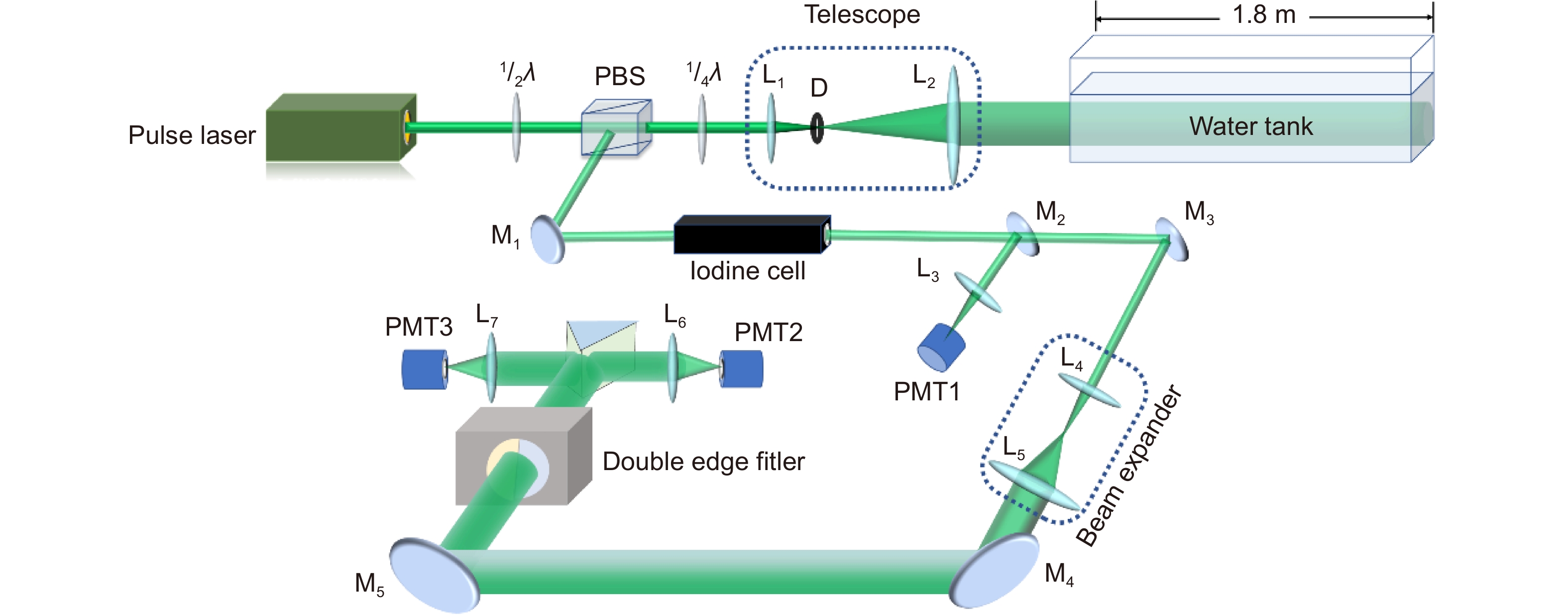
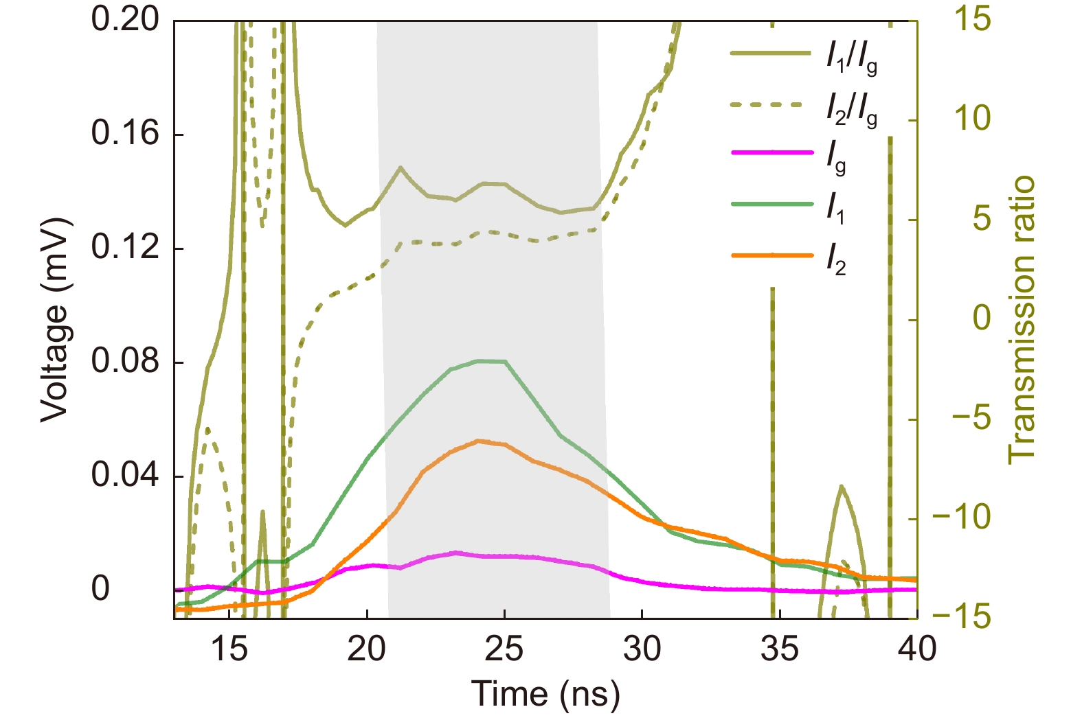
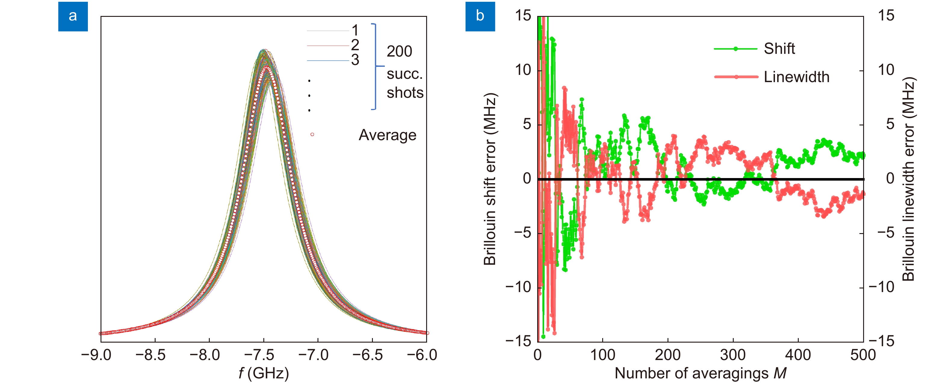
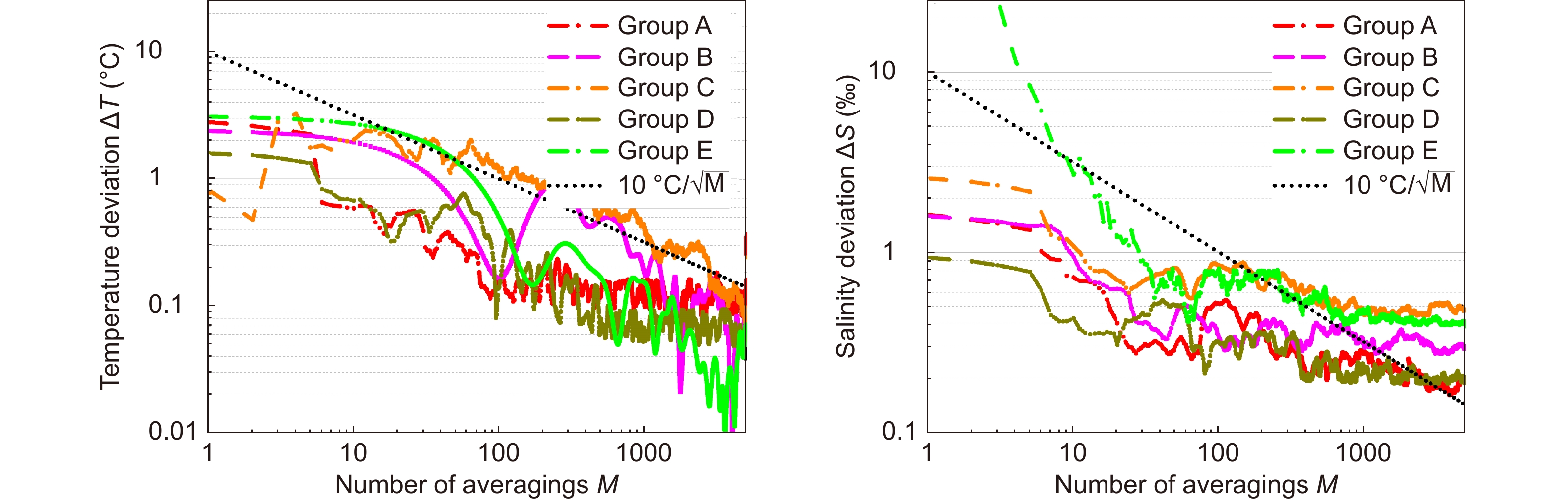
Yang, F., Chen, W., Liang, L. et al. System Design of Ocean Temperature Measurement System Using Brillouin Lidar Based on Dual Iodine Cells. Remote Sensing, 2024, 16(15): 2748.
|
|
| 23. |
Zhang, C., He, Y., Qiao, S. et al. High-sensitivity trace gas detection based on differential Helmholtz photoacoustic cell with dense spot pattern. Photoacoustics, 2024.
|
|
| 24. |
Chen, Y., Liang, T., Qiao, S. et al. Highly sensitive detection of methane based on LITES and H-LITES techniques. Infrared Physics and Technology, 2024.
|
|
| 25. |
Sun, H., Qiao, S., He, Y. et al. Highly sensitive CH4, C2H2and CO simultaneous measurement LITES sensor based on multi-pass cell with overlapped spots pattern and QTFs with low resonant frequency. Optics Express, 2024, 32(16): 28183-28194.
|
|
| 26. |
Zhao, Y., Wang, Y., Liang, K. et al. Underwater Temperature and Salinity Measurement by Rayleigh–Brillouin Spectroscopy Using Fizeau Interferometer and PMT Array. Remote Sensing, 2024, 16(12): 2214.
|
|
| 27. |
Zong, S., Zhang, X., Duan, Z. et al. Research on Laser Dual-Mode Fusion Detection Method of Ship Wake Bubbles. Applied Sciences (Switzerland), 2024, 14(9): 3695.
|
|
| 28. |
Leonov, B.S., Randolph, R.T., Rekhy, A. et al. High-resolution spectroscopy of liquid water with dispersive atomic vapor prism cell. Optics Express, 2024, 32(8): 14847-14859.
|
|
| 29. |
Chen, W., Qiao, S., He, Y. et al. Mid-infrared all-fiber light-induced thermoelastic spectroscopy sensor based on hollow-core anti-resonant fiber. Photoacoustics, 2024.
|
|
| 30. |
Liang, T., Qiao, S., Chen, Y. et al. High-sensitivity methane detection based on QEPAS and H-QEPAS technologies combined with a self-designed 8.7 kHz quartz tuning fork. Photoacoustics, 2024.
|
|
| 31. |
Zong, S., Chen, B., Zhang, X. et al. Laser forward and backward scattering characteristics and experimental study of bubbles in ship wake. Applied Optics, 2024, 63(7): 1795-1810.
|
|
| 32. |
Mao, B., Wu, Y., Cheng, W. et al. Liquid-level sensor based on Michelson interferometer with double hook structure. Microwave and Optical Technology Letters, 2024, 66(3): e34093.
|
|
| 33. |
Shangguan, M., Liao, Z., Guo, Y. Simultaneous sensing profiles of beam attenuation coefficient and volume scattering function at 180° using a single-photon underwater elastic-Raman lidar. Optics Express, 2024, 32(5): 8189-8204.
|
|
| 34. |
Shangguan, M., Yang, Z., Lin, Z. et al. Full-day profiling of a beam attenuation coefficient using a single-photon underwater lidar with a large dynamic measurement range. Optics Letters, 2024, 49(3): 626-629.
|
|
| 35. |
Sun, H., He, Y., Qiao, S. et al. Highly sensitive and real-simultaneous CH4/C2H2 dual-gas LITES sensor based on Lissajous pattern multi-pass cell. Opto-Electronic Science, 2024, 3(11): 240013.
|
|
| 36. |
Guo, Y., Liang, K., Xu, Y. et al. Multiple environmental elements laser remote sensing method based on direct scattering spectrum | [基于直接散射光谱的多环境要素激光遥感方法]. Guangdian Gongcheng/Opto-Electronic Engineering, 2024, 53(1): 240003.
|
|
| 37. |
Liu, Y.H., Qiao, S.D., Fang, C. et al. A highly sensitive LITES sensor based on a multi-pass cell with dense spot pattern and a novel quartz tuning fork with low frequency. Opto-Electronic Advances, 2024, 7(3): 230230.
|
|
| 38. |
Wang, R., He, Y., Qiao, S. et al. Highly sensitive detection of oxygen based on light-induced thermoelastic spectroscopy with a high power diode laser. Infrared Physics and Technology, 2024.
|
|
| 39. |
Zong, S.-G., Yang, S.-P., Zhang, X. et al. Simulation and experiment of weak multi-target laser detection in complex hydrology | [复杂水文微弱多目标激光探测仿真与实验]. Chinese Optics, 2023.
|
|
| 40. |
Zong, S.-G., Zhang, X., Yang, S.-P. et al. Laser backscattering characteristics of ship wake bubble target | [舰船尾流气泡目标激光后向散射特性研究]. Chinese Optics, 2023, 16(6): 1333-1342.
|
|
| 41. |
Shangguan, M., Yang, Z., Lin, Z. et al. Compact Long-Range Single-Photon Underwater Lidar With High Spatial-Temporal Resolution. IEEE Geoscience and Remote Sensing Letters, 2023.
|
|
| 42. |
Wang, Y., Xu, Y., Chen, P. et al. Remote Sensing of Seawater Temperature and Salinity Profiles by the Brillouin Lidar Based on a Fizeau Interferometer and Multichannel Photomultiplier Tube. Sensors, 2023, 23(1): 446.
|
|

 E-mail Alert
 RSS
RSS


 DownLoad: